BBC, (2010). h2g2 The nose, Edited Guide Entry
http://www.bbc.co.uk/dna/h2g2/A27019505 (Accessed 25/5/10)
Cardiac cycle image Figure 7
http://blog.lib.umn.edu/trite001/pstl1082anatomy/2008/09/ (Accessed 25/5/10)
Cleveland Clinic, (2010). Nervous system image http://my.clevelandclinic.org/heart/disorders/electric/syncope.aspx (Accessed 25/5/10)
Faculty of Health, (2010)
http://www.hcc.uce.ac.uk/physiology/circulation02.htm (Accessed 25/5/10)
Hunt, S, (2010). Class notes
Maizels, D. (2003) Oxygen transport and blood flow image
http://www.scientific-art.com/portfolio%20medicine%20pages/respirat.htm (Accessed 25/5/10)
NHS, (2010) Components of blood, The National Blood Service http://www.blood.co.uk/about-blood/components (Accessed 25/5/10)
Probert, M and L, (2010). BRONCHI The Probert Encyclopaedia http://www.probertencyclopaedia.com/cgi-bin/res.pl?keyword=Bronchioles&offset=0 (Accessed 25/5/10)
Red Blood cell image
http://www.owensboro.kctcs.edu/gcaplan/anat2/notes/Notes6%20Blood%20Cells.htm
(Accessed 25/5/10)
SADS, (2003) figure 8
http://www.sads.org.uk/heart_function.htm (Accessed 25/5/10)
Smith N, (2001) Red blood cell; structure and function
http://www.diatronic.co.uk/nds/webpub/red_blood_cell_structure.htm (Accessed 25/5/10)
Think Quest, (2006) Respiratory system http://library.thinkquest.org/2935/Natures_Best/Nat_Best_Low_Level/Respiratory_page.L.html#bronchi (Accessed 25/5/10)
Tutor vista, (2010) Gaseous exchange image
http://www.tutorvista.com/topic/gaseous-oxygen (Accessed 25/5/10)
University of Maryland Medical centre, (2010). Respiratory system
www.umm.edu/respiratory/anatomy.htm (Accessed 25/5/10)
Walmsley, J. (2003). Folens, GCSE P.E for AQA. Folens Publishers, Dunstable.
Wright, D. (2007) Human Physiology and Health. Cambridge Publishing Ltd, 2000. Cambridge.
Friday, 28 May 2010
Relate the structure of the respiratory system to the process of ventilation
 Figure 1
The respiratory system is made up of the
· Nose
· Pharynx
· Larynx
· Trachea
· Bronchi
· Bronchioles
· Alveoli
· Lungs
· Diaphragm
Figure 1
The nose
The nose is made up of bone, cartilage and fatty tissue; it has two holes known as the nostrils. It is separated into two air passages. Goblet cells in the lining of the nose secrete mucus to help trap and remove unwanted antigens. The nose contains tiny hairs known cilia these form the nose lining and keep the mucus moving towards the exit of the nose. Air enters and leaves the respiratory system through the nose.
(BBC, 2009)
Trachea
This is a long tube, around 12 centimetres in length and 2.5 centimetres wide, which is made up of rings of cartilage to keep it open to allow air to flow down it.
Bronchi
Two cartilage-ringed tubes containing smooth muscle tissue, these tubes branch off from the trachea, the right primary bronchus is wider than the left, which makes it more venerable to foreign antigens. These allow air to travel into the lungs, and enter smaller tubes. The bronchi are able to expand during inspiration, to allow the lungs to expand, and contract during expiration as air is exhaled. (Probert, 2010)
Bronchioles
Small thin tree- like network of tubes that contain less cartilage than the bronchi. The bronchioles become smaller than 0.5 mm in diameter, irregularly shaped discs of cartilage support the first few sections, while the latest levels of the tree have no support whatsoever, these sub-divide into the alveolar ducts. By this time air reaches this part all the foreign antigens have been removed and the air travels into the alveoli.
Alveoli
Tiny air sacs that look like a bunch of grapes, they are one cell thick, this helps to speed up gaseous exchange, this action is vital to live. The lung has many millions of alveoli, this gives the lungs a larger surface area for gas exchange. The alveoli are covered in inter linking capillaries through which blood flows. The alveoli diffuse oxygen into the blood stream which bines with the haemoglobin and gets carried round the body, carbon dioxide is then diffused back from the used blood cells into the alveoli and gets breathed out as this is poisonous to the body.
(Think Quest, 2006)
Diaphragm and intercostals muscles
The alveoli are ventilated by the action of the diaphragm and the intercostals muscles.
The diaphragm is a sheet of tendon surrounded by muscles, when relaxed it is positioned into a dome shape by the organs. When the diaphragm muscles contract it becomes flatter. Intercostal muscles are located between the ribs externally and internally. These intercostal muscles allow the ribcage to pivot upwards and outwards, and downwards and inwards. Breathing involves the diaphragm, intercostals muscles and ribcage creating pressure changes. When we breathe in, the external intercostals muscles contract and the rib cage moves upwards and outwards, creating an increase in lung volume and a decrease in pressure. At the same time the diaphragm muscles contract and causes it to move downwards. When we breathe out, internal intercostals muscles contract and the ribcage moves downwards and inwards, causing a decrease in lung volume and an increase in pressure, whilst the diaphragm muscles relax, and the it returns to the dome shape.
(Wright, 2007)
Explain the electrical activity of the heart during a heart beat
 Figure 9
The heart is myogenic (self stimulated), it works by two parts of the nervous system. The sympathetic part of the nervous system accelerates the heart beat, this helps to meet the bodies demands ie when exercising, and the parasympathetic system stops and slows things down.
The rhythmic sequence of contractions (heart beat) is coordinated by the sino atrial (SA), and atrio ventricular (AV) nodes. The upper wall of the right atrium is where the SA node is located and is responsible for sending electrical impulses that initiates atrial contraction. Once the impulse reaches the lower right atrium where the AV node is situated, the impulse remains static for a short period of time, allowing all the blood in the atria to fill the ventricles, before being conducted through several structures which leads to contraction of the ventricles. (Class notes, 2010)
Describe the structure of the heart and explain the cardiac cycle
 figure 8
The heart is a muscular pump that pushes blood around the body, found between the breastbone and the ribs. It is a cardiac muscle and it works involuntary. The heart has for chambers, the atria are the upper chambers, which receive blood, and the ventricles eject blood into the arteries. It has four valves separating the chambers, Tricuspid, separates the right atrium from the right ventricle, Pulmonary valve, separates the right ventricle from the pulmonary artery, Bicuspid valve separates the left atrium from the left ventricle and the Aortic valve separates the right ventricle from the aorta. The heart has three layers, epicardium, gives it a smooth texture, myocardium, responsible for the pumping action, made up of muscle fibres which connect to electrical synapses, and endocardium, inner layer that connect to large blood vessels. The left side of the heart receives oxygenated blood and the right receives deoxygenated blood. (Class notes, 2010)
Cardiac cycle
The cardiac cycle is the sequence of events including contraction and relaxation of the heart. The right side of the heart receives deoxygenated blood from the vena cava, and fills the right atrium, it then flows through the tricuspid valve and into the right ventricle. The tricuspid valve is then forced shut by ventricular pressure, ventricular systole forces the blood up through the pulmonary valve in to the artery, the ventricles are at a diastole stage which makes the pulmonary valve close to prevent back flow. Once the blood reaches the lungs it becomes oxygenated blood. It then gets transported back to the heart via the pulmonary vein and entering the left atrium, from here it goes through the bicuspid valve into the left ventricle. It then gets pumped through the Aortic valve and exits the heart under great pressure out of the aorta which then travels round the body and goes to the cells that require oxygen, the cycle then starts over again. (Class note, 2010)
Describe the structure of the arteries, veins and capillaries and relate this to their function.
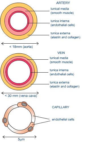 figure 7
Arteries
Arteries transport blood away from the heart; there are two types the pulmonary arteries and systemic arteries. Pulmonary arteries carry blood from the heart to the lungs where the blood picks up oxygen. Systemic arteries deliver blood to the rest of the body.
It has thick muscular and elastic walls as blood is pumped through at a high pressure, from the heart and so that they can expand and contract to push the blood through to the capillaries. They are under sympathetic nervous control that adjusts their diameter to increase or decrease their resistance to the flow of blood. As the arteries get nearer to the tissue they get narrower, these are known as arterioles.
Veins
As blood flows from the capillaries it enters slightly larger vessels know as venules, from here they join the veins. Veins carry deoxygenated blood with the exception of the pulmonary vein back to the heart. The walls are made up of elastic and muscular fibres and are thinner than arteries; it has a larger lumen than the arteries. The veins include valves to ensure the blood does not flow back in the wrong direction.
Capillaries
Capillaries are found at the end of the arterioles, they are the smallest blood vessels in the body; they take the blood through the tissue. They are only one cell thick this helps with diffusion of oxygen and glucose. (Wright, 2007)
Explain the transport of oxygen and carbon dioxide in the blood

figure 6
The red blood cells carry oxygen from the lungs to the rest of the body tissues. A small amount of oxygen (1.5 %) is carried in the plasma as a dissolved gas. Most oxygen (98.5 %) carried in the blood is bound to the protein haemoglobin in red blood cells. A fully saturated oxyhaemoglobin has four oxygen molecules attached. Oxygen is transported by blood to enable us to respire which is essential to live.
Carbon dioxide is more soluble in the blood than oxygen, about 5% is transported unchanged. Carbon dioxide combines with haemoglobin to form carbaminohaemoglobin. 10% of carbon dioxide binds to amino groups on the polypeptide chains of haemoglobin and plasma proteins. Carbon dioxide enters red blood cells in the tissue capillaries where it combines with water to form carbonic acid. This is normally a slow reaction, but the RBC is greatly accelerated by an enzyme called carbonic hydrase. (Smith, 2002)
The heart acts as a central pump that keeps the blood moving around the body. When air is inhaled, it enters the lungs and diffuses through the alveoli; the oxygen binds with the haemoglobin of the red blood cells to form oxyhaemoglobin. The blood then enters the pulmonary circulation system, where it gets pumped round to cells that require energy for cellular respiration. Oxygen diffuses out of the red blood cells and carbon dioxide diffuses in making the blood deoxygenated, it then travels back to the lungs and diffuses into the alveoli and is exhaled as the waste product carbon dioxide.
(Class notes, 2010)
Describe the structure of a red blood cell and explain how this relates to its function

Red blood cells have a unique shape for their function.
· Flattened, biconcave disc shape
· Don’t have a nuclei or mitochondria
· In adulthood they are produced in the bone marrow
· They live approximately 120 days
· They transport oxygen around the body
· Red colour due to haemoglobin.
(Smith, 2001)
Red blood cells are thinner in the centre and thicker around the edges. They are very flexible with the ability to twist and bend through the blood vessels, as these narrow and widen throughout the body. It function is to carry oxygen around the body to all the tissues from the lungs, the red blood cells than transport the carbon dioxide from the cells back to the lungs. The biconcave shape increases the cell's surface area and facilitates diffusion of oxygen and carbon dioxide into or out of the cell. With the red blood cell not containing a nuclei it helps contribute to increased haemoglobin content and gas-carrying capacity. (Walmsley, 2003)
Figure 5
Subscribe to:
Comments (Atom)